• Surajmal Vihar, Delhi | Indirapuram, Ghaziabad
  • S.V.D - Mon-Sat 9AM to 8PM | Sun: 9AM to 4PM || I.G - All Days 10AM - 8PM

Diabetic Retinopathy Treatment in Delhi NCR

Diabetes Mellitus is a condition that impairs the body’s ability to use and store sugar (glucose). Sugar is excreted in the urine and the blood sugar is abnormally high. It causes changes in small blood vessels in various organs of the body. Diabetes can cause various changes in the eye as well, particularly in the retina. Diabetic eye disease can cause severe vision loss or even blindness. 

Learn About Diabetic Retinopathy

04
03

How The Eye Works

The eye is like a camera in which lenses focus the picture on a light sensitive film. In the human eye the transparent cornea and lens focus images on the retina, a thin light sensitive film which receives light and changes it into electrical signals which are then transmitted to the brain by the optic nerve.

Just in front of lens lies the iris (‘colored portion of the eye’) with a central opening – the pupil. This is just like the shutter or diaphragm aperture of the camera and helps regulate the amount of light entering the eye. The sclera (‘white of the eye’) is the protective outer coat of the eye. Between the sclera and the retina lies the choroid, which has a chiefly nutritive function.

The space between the lens and the retina is filled with a clear jelly called the vitreous body. 

How The Eye Works
Retina Treatment Delhi

The Retina

The retina can be divided into two regions: the central (macula) and the peripheral. The macula is a very small area in the centre of the retina upon which light rays are focused by the cornea and the lens of the eye.

The macula being the most sensitive portion of the retina is responsible for acute (sharp) vision, – that is for reading, writing, threading a needle and other fine tasks. The periphery of the retina gives us vision to the side, which is called “peripheral vision”. This is what we refer to when we say, “i saw something from the corner of my eye.”

What Is Diabetic Retinopathy?

When the retina is affected by diabetes, weakened blood vessels may leak fluid or blood, causing damage to the retina. This is called diabetic retinopathy.

Diabetic Retinopathy in Delhi
Background Retinopathy

There are two forms of diabetic retinopathy. In background retinopathy, blood vessels within the retina become abnormally permeable and allow substances like fluid and lipid to leak out.

This results in water logging or edema of retinal tissue and deposition of yellowish material called exudates. If the leaking fluid collects in the macula (the central part of the retina responsible for reading vision and other fine tasks), vision gets affected, often to a marked degree.

Proliferative Retinopathy

The second form is proliferative retinopathy. Abnormal fragile ‘new’ blood vessels grow on the surface of the retina or optic nerve and sometimes into the vitreous cavity. These fragile vessels may rupture and bleed into the normally clear vitreous gel, blocking light from reaching the retina.

Subsequent scar formation in the vitreous may pull on the retina, detaching it from the back of the eye (traction retinal detachment). Severe loss of sight, blindness, and even painful glaucoma can result from these conditions.

What Are The Symptoms?

Please note that every person with diabetes need not have diabetic retinopathy. Conversely an eye with marked changes of diabetic retinopathy can have good vision and be totally free of symptoms.

Regular Eye Checkup

Hence it is important for all diabetics to undergo regular eye check-up including retinal examination through dilated pupils especially for people who have been diabetic for a number of years. It is also true that diabetes is often detected in a person, when some changes of retinopathy are seen on routine examination of the eye.

Reduced central vision can occur if the macula gets edematous (swollen). Black spots (floaters) and cobwebs of sudden onset often point to a minor bleed inside the eye. Sudden total loss of vision may occur due to a large bleed into the vitreous

Investigations For Diabetic Retinopathy

If diabetic retinopathy is noted, color photographs of the retina may be taken and fluorescein angiography performed. This involves dilating the pupils and injection of a fluorescent dye into a vein in the arm.

Photographs of the retina are taken rapidly as the dye passes through the retinal blood vessels. This test helps in determining if laser photocoagulation treatment is necessary. If treatment is to be done, it helps in identifying what structures and areas need treatment with laser.

Optical coherence tomography (OCT), which is newer non-invasive diagnostic modality provides a cross-sectional view of the retina and helps in quantifying the amount and type of swelling and guides the treatment. 

Treatment Of Diabetic Retinopathy

Photocoagulation involves the use of a laser beam to seal leaking blood vessels and prevent growth of abnormal blood vessels. This procedure does not require hospitalization. In background retinopathy, if blood vessels are leaking fluid into the macula, laser treatment stops the leakage and may improve or stabilize vision.

Proliferative Retinopathy

In proliferative retinopathy, laser treatment may involve one or more sessions depending on the type and severity of retinopathy. Laser treatment significantly reduces the chances of severe visual loss by destroying the abnormal blood vessels and preventing growth of more such vessels.

Vision may improve or stabilize within several weeks to a year. It is important to remember that laser treatment is not a one-time procedure. Regular follow up is extremely important. Your doctor will tell you when to return for a check-up.

Recently, along with laser treatment, certain medication when injected into the eye or just outside the eye has shown encouraging results. These medicines are however to be used cautiously and judiciously.

Vitrectomy

If the vitreous is too clouded with blood or there is traction retinal detachment, laser treatment will not work. In this situation, a surgical procedure called vitrectomy needs to be performed. In this operation, opaque vitreous gel is removed from within the eye by a special instrument that simultaneously sucks and cuts the vitreous. 

Prevention Of Diabetic Retinopathy

LOSS OF VISION FROM DIABETIC RETINOPATHY IS LARGELY PREVENTABLE.

Early detection of diabetic retinopathy is the best protection against sight loss. This is possible by having a complete eye examination including retina check-up once a year or more frequently if advised. In most cases the ophthalmologist can then begin treatment before sight is affected.

Excellent control of diabetes and associated conditions like hypertension, increased blood lipids & cholesterol and renal (kidney) disease, is strongly recommended. However, good control in itself does not guarantee freedom from diabetic retinopathy.

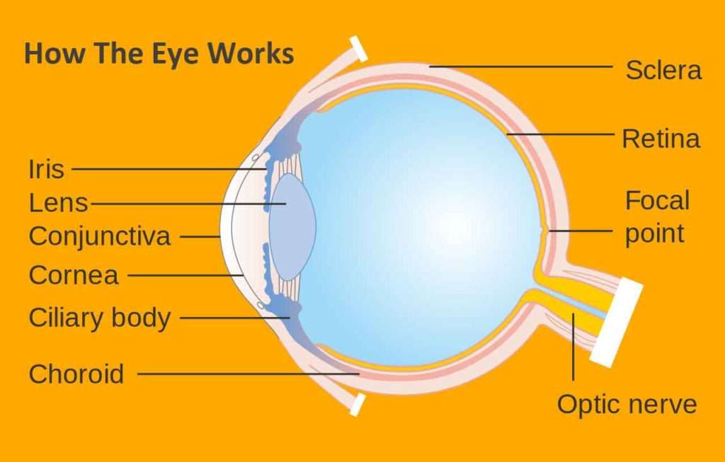
Schedule Your Eye Care Appointment

Submitting your appointment request is the initial step towards personalized and timely eye care.

Our team will contact you within 24 hours to confirm your appointment. For immediate assistance, please call us directly.

Loading...